Anatomy and Physiology of the Human Heart


Â
The heart is a muscular pump, cone shaped, hollow organ that lies in the chest cavity, the apex inclining towards the left cavity. It is divided into four areas, the upper right and left atria and the lower right and left ventricles.
A muscular wall called the septum down the centre separates oxygenated from deoxygenated blood. The hearts purpose is to circulate blood throughout the body.
The wall of the heart has three layers the Inner layer (endocardium).
The middle layer (myocardium).
The outer layer (pericardium).
The action on the left side is to receive blood from the lungs and to force it around the body. The action on the right side forces blood into the lungs to be oxygenated.
Valves are found between the atria on upper part and ventricles on lower part.
Cardiac Cycle
There are three stages to the event of a heartbeat.
- Blood enters the heart, the atria and ventricles are both relaxed or DIASTOLE. Blood enters the atria while all the valves are closed.
- Blood is pumped from the upper atria to the lower ventricles. Electrical impulses from the pacemaker cause the atria to contract ATRIAL SYSTOLE. Blood is pumped to the ventricles. The tricuspid and bicuspid valves open. The vena cava and pulmonary veins close to stop blood entering the atria.
Blood leaves the heart and the atria relax. Impulses from the av node cause the ventricles to contract. This is called ventricular systole. Blood id forced out of the heart into the pulmonary artery and the aorta. Pressure forces the semilunar valves to open. Pressure closes the tricuspid and bicuspid valves. Ventricles relax again. Semilunar valves close which prevents blood from flowing back into the heart or ventricles. The vena cava and pulmonary veins open and the cycle starts again.
Blood pressure
Blood pressure is the power exerted by the blood against the blood vessels walls , and and the arteries, while it becomes lower in the veins and capillaries.
Blood pressure is read with a sphygomomanometre.
SYSTOLIC:
Heart is contracting blood pressure reaches its highest point.
DIASTOLIC:
Pressure reaches its lowest level when the heart IS relaxing.
High Blood Pressure or Hypertension.
Causes
Narrowing of the arteries, Kidney disease, smoking. Diet, hereditary factors including stress and medication. High blood pressure is maintained at a high level over a period of time.
Symptoms
Heart attack, Stroke, Kidney complaints, Angina.
Low Blood Pressure or Hypotension.
Causes
Low blood pressure is maintained over a period of time. It can be shock or an underactive Adrenal glands, or hereditary factors.
Symptoms
Fainting and dizziness.
- CARDIAC OUTPUT
Volume of blood pumped out of the heart.
When cardiac output increases blood pressure increases.
- RESISTANCE OFFERED BY ARTERIOLES (small arteries).
Narrowing of blood vessels can result from contraction of the muscular wall of the vessels. The greater the narrowing the higher the blood pressure.
- TOTAL BLOOD VOLUME
Blood pressure is lowered if the amount of circulating blood is reduced
- VISCOSITY OF BLOOD
The lower the viscosity the lower the blood pressure.
- ELASTICITY OF ARTERY WALLS
   When arteries harden there is a loss of elasticity and blood pressure is raised.
Structure of Arteries Veins and Capillaries
|
Characteristics of capillaries |
Characteristics of veins |
Characteristics of arteries |
|
Distribute oxygen & nutrients to all cells of body |
Veins transport blood to Heart |
Arteries transport blood from the heart |
|
Transport Carbon Dioxide & other waste away from cells. |
Transport deoxygenated blood except pulmonary |
Arteries transport oxygenated blood except the pulmonary. |
|
Capillaries are smallest blood vessel. One cell thick, |
Veins carry a high concentration of urea & waste, |
Arteries have an abundance of nutrients. |
|
Capillaries have thin walls. |
Not as muscular & elastic compared to arteries. |
Elastic Walls. Muscular. |
|
The fluid, mostly water & nutrients filters out of walls & bathes body tissue. |
The Lumen, i.e. the passage is large. |
The Lumen i.e. the passage is small. |
|
Valves stop the blood flowing back. Pumped by skeletal muscles. |
Arteries are pumped by heart & muscle tissue in the artery wall. |
|
|
Blood under low Pressure |
Blood under high pressure. |
|
Skeletal muscle |
Smooth muscle |
Cardiac muscle |
|
Voluntary |
Involuntary |
Involuntary |
|
Stripped With Protein Bands. |
Non Stripped. Or Non-Striated. |
Is The Pump To Power The Heart |
|
Joined Onto The Bones. |
We Have No Control Over Them. |
Only Found In The Heart |
|
Consciously Control |
Found In The Digestive System, Respiratory System And The Genito Urinary System. |
Outer Fibres Are Striated Or Striped |
|
Numerous Nuclei |
Automatically Work. |
Only One Nucleus. |
|
Made Of Fibres That Form A Group Of Cells. |
One Nucleus |
Looks Like Skeletal Muscle. |
|
Largest Cells In The Body |
Spindle Shape With No Distinct Membrane. |
|
|
Sheath On Outer Muscle |
Regulate The Flow Of Blood In Arteries. |
|
|
Fibres Form Into Bundles And Go In The Same Direction. |
Moves Your Dinner Along Through Your Gastrointestinal Tract. |
|
|
Regulate The Flow Of Air Through The Lungs. |
||
|
Help Deliver Babies From The Uterus. |
How Muscles and Skeleton work together to create movement
- A muscle needs to pass over a joint to create movement.
- Muscles are connected to bones by tendon.
- Tendons pull on a bone when a muscle contracts and helps it move.
- Usually, muscles work in antagonistic pairs. In each pair there is a relaxing muscle and a controlling muscle
- Antagonistic muscles must contract and relax equally to ensure a non-jerky smooth movement. An example is the biceps muscle on top of the humerus. The arm is moved upwards. At the same time the triceps is relaxed.
- Body moves when the muscles contract and produce movement in the joints of the skeleton,
- Muscles stabilise the joints,
- Muscles maintain posture control.
- Muscles aid in temperature control e.g., shivering.
- Axial muscles Skeletal Muscles of the trunk or head e.g. trapezius muscle.
- Appendicular Muscles Skeletal muscles of the limbs e.g. biceps triceps. These two muscles contract and relax equally to ensure a smooth, non-jerky movement.
Â
Composition of bone
- Bones are living tissue. They contain Osteoblasts which are responsible for making the collagen rich substance osteoid, which is key in building bone.
- They compose of cells called Osteoclasts that maintain the bone structure. The cells travel around the the bone to areas in need of resorption.
- They compose of compact bone which accounts for 80% of the body’s bone mass.
- They compose of cancellous bone makes up 20% of the bodies bone mass. It has a honeycomb structure.
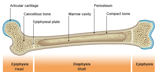
STRUCTURE OF A LONG BONE
External structure
Long bones fornexamole the femur in the leg are enclosed in a membrane called the periosteum. This membrane contains blood vessels and nerves. The long shaft of a bine is called the epiphysis.
Internal Structure
- Compact bone
- Spongy bone
- Medullary cavity
Compact bone
Is mostly found in the shaft or diaphysis of a bone. It is also found around the end or epiphysys of a bone. When under a microscope bones are full of holes.
Haversian Canal:
These are canals that run lengthways through compact bone. They contain blood capillaries, and nerves.
Cancellous or Spongy bone:
These bones are found at the end of long bones and are found in flat and irregular bones. It is spongy.
Medullary Cavity:
The red and yellow bone marrow is stored here.
Functions of the Skeleton
- Allows movement as joints are formed between the bones to allow the movement of the body.
- Provides attachment for the muscles which move the joints thus moving the body.
- Supports the body and gives it shape as all the other parts of the body are soft and cannot stand up.
- It protects the delicate organs e.g. Skull as it protects the brain, the rib cage. The sternum protect the Heart and lungs.
With the aid of vitamin K calcium salt and phosphorus is stored in your body.
Different Types of Joints
FREELY MOVEABLE SYNOVIAL JOINTS
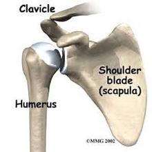
Shoulder joint is called a ball and socket joint. It is the most moveable joint. It allows movement in many directions. The rounded head of one bone fits into a socket or cavity in another bone.
Immoveable or fixed fibrous joints
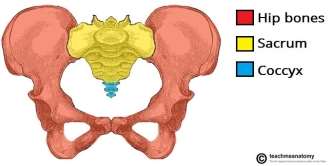
Innominate or pelvic girdle bone has no movement. There is fibrous tissue between the ends of the bones.
Slightly moveable joints cartilaginous
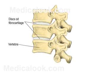
When two bones come together with a little cartilage in between. Some examples would be – the joints between all of the vertebrae in the spine. These bones are around the discs, which are made of cartilage.
|
Freely Moveable Synovial Joints |
Immoveable or Fixed Fibrous Joints |
Slightly Moveable Cartilaginous Joints |
|
Ball & Socket Joint |
Pelvic Girdle or Innominate Bone |
Joints Between Each Vertebrae in The Spine |
|
Hinge Joint |
Sutures in Skull |
Symphysis Pubis in The Pelvis. |
|
Gliding Joint |
Sacroiliac Joint in The Pelvis |
|
|
Pivot Joint |
||
|
Saddle Joint |
Body Movement of How the Skeletal and Muscular System Connect
In the skeletal muscles, a muscle needs to pass over a joint to create movement. Tendons connects muscles to bones. When the muscle contracts, the tendon pulls on the bone and causes it to move. Most of the time muscles work in antagonistic pairs. Each pair consists of a contracting muscle and a relaxing muscle. These two muscles must contract and relax equally to ensure a smooth, non-jerky movement.
Therefore, the muscles contract and allow movement in the joints of the skeleton causing the body to move. The muscles stabilise the joints and ensure posture control. The axial muscles are the muscles of the trunk or head. Then the appendicular muscles are the skeletal muscles of the limbs, e.g. Biceps, triceps.

- Epidermis:
Structure
Consists of five layers on the upper portion of the skin. Cells in the bottom are living and carry on moving up through the layers until they die.
Function
To protect the skin.
- Dermis:
Structure
Lies beneath the epidermis.
The papillary layer is wavy tissue. The waste upward projections are called dermal papillae. They contain blood and lymph capillaries and nerve endings.
The reticular layer contains the main components of the skin. It is dense and fibrous.
Function
The papillary layer increase surface area of reproductive cells and provide living layers of epidermis with vessels which supply nourishment and remove cellular waste.
The reticular layer protects and repairs injured tissue.
Collagen gives it strength.
Elastin allows the skin to stretch easily but quickly regain its shape.
- Subcutaneous layer:
Structure
Lies beneath the dermis has cells called lipocytes which produce lipids which are the fat cells from which subcutaneous tissue is formed.
Function
Cushions muscles, bones and internal organs against shocks and blows.
- Sudoriferous glands:
Structure
Found in the dermis.
Eccrine glands
Found all over the body, numerous on the palms of hands and the soles of the feet. They produce sweat through a sweat pore.
Aprocrine glands are found in the armpits, nipples and anal and genital areas open into hair follicles and produce a thicker secretion.
Function
Eccrine glands help regulate body temperature by producing sweat which evaporates off the skins surface and cools it down when it is hot.
Apocrine glands are under nervous control and respond to emotional, psychological and sexual stimuli.
- Hair follicles:
Structure
Found all over the body except the palms of hands and soles of the feet. It is a sac like structure which contain hairs. The base of the hair degenerates and rebuilds during the cycle of hair growth and replacement. It contains a dermal papilla which supplies blood to the base of the hair.
The follicle opens at the skins surface at a follicular pore.
Function
Hair follicles produce and contain hairs during their life cycle. They provide nourishment for the hairs.
- Hairs:
Structure
Found in the follicle in the dermis. They do not grow on lips, palms of hands, or soles of feet.
The hair above the skin is called the shaft. The portion lying in the follicle is called the root.
The enlarged base of the root surrounding the papilla is called the bulb.
Hair is made of protein keratin.
Function
Hair protects against friction and damage from external environment.
Hair is a sexual characteristic.
- Sebaceous glands:
Structure
Found in the dermis and produce sebum which pass through a duct and up the hair follicle and through the skin through a follicular pore.
Function
Sebum lubricates the skin and hair and combines with sweat to form the protective acid mantle of the skin.
It also retains natural moisture in the skin and provides insulation.
- Blood Vessels:
structure
Arteries carry oxygenated blood. Blood is pumped all around the body in arteries.
Veins carry deoxygenated blood. Their walls have valves which stops blood from flowing backwards.
Capillaries are fine vessels and made of a single layer of cells. Some materials can pass in and out through the thin walls of the capillaries.
Function
Arteries carry oxygen and nutrients to the skin via capillaries.
Veins remove waste products.
The surface capillaries help to regulate body temperature.
Vessels dilate and heat is lost through the skin.
When the body is cold, the vessels contract and heat is retained.
- Nerves:
structure
Found in the dermis and subcutaneous tissue.
Sympathetic nerves supply blood vessels, sweat glands and the arrector pili muscle.
Nerves respond to heat, cold, pain, pressure and touch.
Function
Nerve stimulation causes a reaction which triggers an appropriate response from the body.
SECTION D
Examples of Viral Bacterial, Fungal Skin Diseases:
|
Viral skin disease |
Bacterial skin disease |
Fungal skin disease |
|
Chickenpox |
Cellulitis |
Athletes Foot. Tinea Pedis |
|
Mumps |
Impetigo |
Ringworm |
|
Hepatitis B Virus |
Folliculitis |
Jock Itch |
|
German Measles / Rubella |
Furuncle |
Fungal Nail Infection Onychomycosis |
Relationship Between Skin & The Nervous System
- Sensory nerve endings are situated in the skin and give us the sensation of touch.
- The nerve endings or “receptors” are specially shaped and positioned to respond to a range of different stimuli. We can distinguish heat, cold, and pain, as well as differences between light and deep pressure.
- Motor nerve endings supply the muscles that make facial expressions and move the eyes, neck, and lower jaw.
- Made up of white or grey nerve fibres which end in sensory nerve endings.
- Nerve stimulation causes a reaction which sets off an appropriate response from the body.
- The skin also is important in helping to switch your body temperature. If you are too hot or too cold, messages are sent from the brain to the skin. The skin uses 3 methods to increase or decrease heat loss from the body’s surface – these are hairs on the skin trap heat if when standing up, and less if they are lying flat; glands under the skin secrete sweat onto the surface of the skin in order to increase heat loss by evaporation if the body is too hot; capillaries near the surface can open when your body needs to cool off and close when you need to conserve heat.
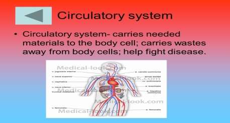
Relationship Between the Skin and The Circulatory System.
- The circulatory system through the help of arteries, veins and small capillaries transport blood, nutrients and oxygen to the skin. This is done by the help of the pulmonary circulation and the systemic circulation.
- Arteries carry a lot of nutrients via the circulation system to the skin.
- Veins carry a lot of waste products like urea to the skin.
- The skin protects the body living tissues and the organs.
- It protects against the invasion of infection
.
- It protects from dehydration.
- The circulatory system protects the body against changes in temperature. The skin has nerve endings called thermoreceptors which detect hot a and cold. These receptors interact with a cluster of nerves at the centre of the brain called the hypothalamus.
If you become too hot or too cold, your brain sends nerve impulses to the skin, which has three ways to to decreases or to increase heat loss from the surface of the body.
BIBLIOGRAPHY:
Biology Plus Leaving Cert by Michael O Callaghan Edco.
A Practical Guide to Beauty Therapy Level 2 By Janet Simms.
Internet:
“Teachpe.Com – Free Resource For Physical Education And Sports Coaching”. Teachpe.com. N.p., 2017. Web. 26 Jan. 2017.
“Sciencedirect.Com | Science, Health And Medical Journals, Full Text Articles And Books.”. Sciencedirect.com. N.p., 2017. Web. 27 Jan. 2017.
“Innerbody.Com | Your Interactive Guide To Human Anatomy”. Innerbody. N.p., 2017. Web. 30 Jan. 2017.