Cephalometric Assessment of the Sella Turcica in Children
Keywords: radiographs, sella turcica, children, shapes
ABSTRACT:
Background: On a lateral skull radiograph the image of sella turcica is U shaped. A deviation from normal size and shape of sella turcica can be an indication of a pathological condition of the pituitary gland. The center of sella turcica is routinely used as a cephalometric landmark to act as a reference point for evaluating spatial position of both jaws as they relate to the cranial base. One of the most commonly used cranial landmarks for cephalometric tracing is sella point. The morphology and size of sella turcica is of importance because within its center lies sella point which helps in evaluation of craniofacial morphology.
Methods: All lateral skull radiographs taken in the department of radiology were retrieved for the study. The total numbers of radiographs were two hundred and fifty (250) and only 162 of these satisfied the inclusion criteria. Radiographs were mounted on the viewing box and variants of the anatomical shapes of the sella turcica were studied and classified.
Results: The predominant shape of sella in the Nigerian children studied was round, and the difference in frequency of round shape sella and that of oval type is highly statistically significant (p< 0.001). The commonest type of sella turcica floor in the Nigerian children studied was concave and the difference in frequency of concave shape sella floor and that of flat type is highly statistically significant (p <0.001). In both the various anatomical shapes of the sella turcica and the type of floor of the sella turcica in relation to sex of the Nigerian children studied, the difference in frequency of males and females is highly statistically significant (p < 0.001).
Conclusion: The prevalence and the relative frequencies of the normal variants of the anatomical shapes of the sella turcica of male Nigerian children differ significantly from those of their female counterparts further studies on a large scale are needed to corroborate our findings.
Keywords: Sella turcica, shapes, radiographs, children.
INTRODUCTION
The sella turcica is an important anatomical structure for cephalometric assessment because of its central landmark; it lies on the intracranial surface of the body of the sphenoid and consists of a central pituitary fossa bounded anteriorly by the tuberculum sellae and posteriorly by the dorsum sellae.1
The center of sella turcica is routinely used as a cephalometric landmark to act as a reference point for evaluating spatial position of both jaws as they relate to the cranial base. One of the most commonly used cranial landmarks for cephalometric tracing is sella point. The morphology and size of sella turcica is of importance because within its center lies sella point which helps in evaluation of craniofacial morphology.2
On a lateral skull radiograph the image of sella turcica is U shaped. A deviation from normal size and shape of sella turcica can be an indication of a pathological condition of the pituitary gland.3
Jones reported that the anatomy of the sella turcica is variable in size and shape and classified sella into three types; round, oval and flat.4
Gorden and Bell examined radiographs of children of age ranging from 1 year to 12 years, classifying the sella into 3 shapes; the circular, the oval or the flat/saucer shaped. Gordon & Bell concluded that most of the subjects in their study had either circular or oval shaped sella turcica.5
The sella turcica have been described based on the contour of the sella turcica, the angle made by the contour of tuberculum sella, the contour of the anterior and posterior clinoid processes and the fusion of both the processes termed as ‘ sella turcica bridge’.6,7,8
In a recent study by Axelsson shape of the sella turcica was divided into six main types; normal sella turcica, oblique anterior wall, double–contoured sella, sella turcica bridge, irregularity (notching) in the posterior part of the sella and pyramidal shape of the dorsum sellae.9 The normal variation of sella turcica was seen in 2/3rd of the subjects while the remaining subjects showed dysmorphological appearance.10 The variation of the sella turcica morphology apart from normal can be misleading since it may be present in normal patients as well as medically compromised patients as seen in spina bifida or craniofacial deviation.11
In a study by Silverman, of children radiographs aged between 1 month and 18 years. He reported that pituitary fossa of males tend to be larger than the females from 1 to 13 years of age. Because of the pubertal growth spurt which occurs 2 years earlier than males, a significant increase in size of the sella turcica occurs from 11 to 15 years of age in the females. 12 Thereafter the pubertal growth spurt in males occurs about 2-3 years later than females resulting in approximately equalization in sella area in both genders. 12
In children, 70% of sella are round, in adults only 24.4% are round, and whereas 58% are oval and 17.2% are flat13. Haas reported that till the age of 17 the mean size area of sella turcica in males was more, however after age of 17years the sella turcica area in females is slightly larger compared to males.14 Plain film radiographs have a relatively high sensitivity for detecting sella change at between 67% and 77% of positive findings and clinicians should be suspicious when any of the sella turcica dimensions exceed the upper limits of normal.15
The sella floor is recognized in the postero-anterior view of the skull film in over 90% of cases and in 100% of cases using tomography.16, 17
Bruneton et al. studied 200 radiographs of normal adult from North American and noted the percentage of variants of each anatomical feature on both standard radiograph and tomography. In this series the floor was concave in 58% of subjects, flat in 32.5% and convex in 9.5%. Thus, the floor of the sella turcica which in most cases is concave may be, flat or even convex19.
The objective of this study was to describe the shape and floor of the sella turcica in children.
MATERIALS AND METHODS
Study Design:
All available lateral skull radiographs of Nigeria children over a 3-year period from 20013 to 2014 were retrieved from the radiology department for the study.
This study was a cross sectional comparative study. All the radiographs were ascertained to have been taken by a trained radiographer in a standardized condition/manner (focus to film distance target to film distance (FFD/TFD) OF 40 inches (100cm) (Stewart and bushing, 1980).
All the lateral skull radiographs included in the study showed clear reproduction of Sella turcica and were of patients 15 years old or below.
The total number of radiographs was two hundred and fifty (250) and only 162 of these satisfied the inclusion criteria. Only radiograph interpreted by experienced radiologist were studied.
Inclusion and Exclusion Criteria
Perfect superimposition of the crinoids processes, to rule out tilting of the skull during positioning of the patient, clear visualization and recognition of the dorsum sellae and tuberculum sellae, distinct sella turcica floor so that the shape of the fossa and its floor could be classified. Syndromic patients or patients with major illness were not included. All the lateral skull radiographs were taken by the same trained operator (Radiographer) using GE Haulum medical system X-ray machine model No.2226519.
Description Techniques:
Radiographs were mounted on the viewing boxes and variants of the anatomical shapes of the sella turcica were studied and classified according to the method adopted by Bruneton et al, 1979 and Zagga et al, 2008
Statistical Method:
Data was initially sorted out manually and tabulated and then entered into the computer Microsoft Excel and Minitab 13.1 statistical package chi-square was use for comparison of proportion. Statistically significant in comparison to other values was set at p<0.001.
RESULTS
A total of 162 subjects were involved in the study. Of these number 106 (65%) were males and 56 (35%) were females (m:f ratio 9:1). The result obtained are summarized in tables 1-4
Table 1: Showing the various Anatomical Shapes of the Turcica
Shape of the fossa Frequency Percentage
Oval 48 29.6%Round 114 70.4%
Total 162 100%
The various anatomical shapes of the sella turcica of Nigerian children seen in the study are shown in table 1. The predominant shape of sella in the Nigerian children studied is round (Figure 1) and the difference in frequency of round shaped sella and that of oval (figure 2)type is highly statistically significant (x2=257.1579;df=1;p 0.001)X2=128.1; df =1; p 0.001
Table 2: Table Showing the various Anatomical Shapes of the Sella Turcica in Relation to Sex.
|
Shape of the floor |
Frequency |
Total |
|||
|
Males (Number) |
Percentage (%) |
Females (Number) |
Percentage (%) |
||
|
Oval |
78 |
73.6 |
36 |
64 |
114 |
|
Round |
28 |
26.4 |
20 |
35.7 |
48 |
|
Total |
106 |
100 |
56 |
100 |
162 |
X2=28.0000, df=1; p 0.001
With regards to the various anatomical shapes of the sella turcica (round and oval) in relation to the sex of the Nigerian children, this study revealed males to be predominant for each of the two shapes of the sella turcica as shown in table 2. The difference in frequency of male and female Nigerian children is highly statistically significant (x2=57, 0000; df=1;p0.001)
Table 3: Table Showing the Types of Sella Turcica Floor seen in this Study.
Shape of the Sella Floor Frequency Percentage
Concave 130 80%
Flat 32 20%
Total 162 100%
X2=180.0263; df=1; p 0.001
Table 3 shows the difference shapes of the floor of the sella turcica. The commonest type of sella floor in Nigerian children studied is concave (Figure 1) and the difference in frequency of concave shaped sella floor and that of flat (Figure 2) type is highly statistically significant (x2=180.0263; df=1; p 0.001)
Table 4: Table Showing the Types of Sella Turcica Floor in Relation to Sex.
|
Shape of the Fossa Floor |
Frequency |
Total |
|||
|
Males (Number) |
Percentage (%) |
Females (Number) |
Percentage (%) |
||
|
Concave |
110 |
84.6 |
20 |
62.5 |
130 |
|
Flat |
20 |
15.4 |
12 |
37.5 |
32 |
|
Total |
130 |
100 |
32 |
100 |
162 |
X2=570000; df=1; p0.001
Table 4 shows the types of sella turcica floor (concave and flat) in relation to sex of the Nigerian children studied. It shows that male Nigerian children predominated for each type of sella turcica floor. The difference in frequency of male and female children is highly statistically significant. (x2=570000, df=1,p 0.001).
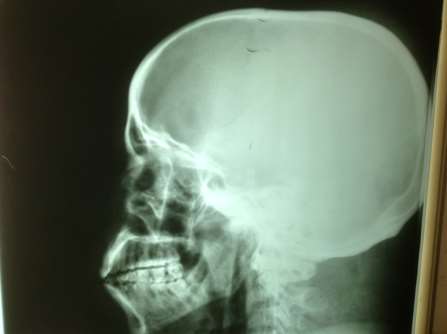

Figure 1: An image of the lateral radiograph of a 13 year old male child showing a round type of sella tucica with a concave floor.

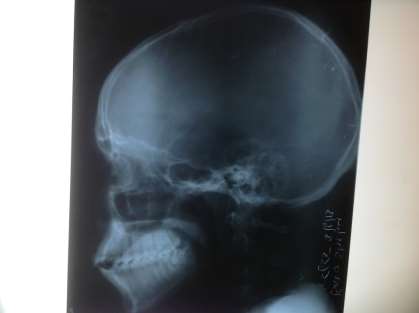

Figure 2: An image of the lateral radiograph of a 3 year old female child showing an oval type of the sella turcica with a more or less flat floor.
DISSCUSION
The dental profession can play an important role in the detection of skull lesions. Orthodontists, in particular, routinely take lateral skull radiographs as part of the process of orthodontic diagnosis, treatment planning, and assessment of therapeutic results.21 Hence they may be the first to observe an abnormality in the sellar region of the cranium. This initial diagnosis by an orthodontist and subsequent investigation and evaluation by an endocrinologist or neurosurgeon might sometimes be lifesaving to the patient. 21
Gorden and Bell examined radiographs of children and classify the sella into 3 shapes; the circular, the oval or the flat/saucer shaped. There conclusion was that most of the subjects had either circular or oval shaped sella turcica5. Similar finding was reported by Jones et al,. When compare to the current study two types of shapes of sella turcica (round and oval) were observed. Although Jones et al, and Gorden and Bell 1922 did not report the percentage prevalence of each of the anatomical type of sella turcica. We found that 70.4% of the sella to be the round type to be in our study. However, this study compares favorably with that of Meschan, who reported that 70% of sella turcica in children as round.
The two type of the sella turcica floor observed in this study have also been reported by Bruneton et al., In both series concave sella floor is commoner19. In this study, the Prevalence of concave type of sella turcica floor was 80%, which is higher than the 58% reported by bruneton et al, 1979. Flat type of sella turcica floor appeared to be less common. The prevalence (20%) of flat type of sella turcica floor found in this study is lower than that (32.5%) reported on Caucasian by Bruneton et al.
Ahsan et al reported variation in the shape of the sella in 34% of the subjects in their study: an irregular dorsum sella was found in 16.7 %. A pyramidal shape was present in 7.7%, double contour-sella was found in 5.5%, an oblique anterior wall was found in 4% while sella turcica bridge was found in none of the patients22. This differs from the findings in our study where two types of shapes of sella turcica (round and oval) were observed; ethnic factor could account for this differences in the shaped of sella turcica.
Although CT scan and MRI have replaced plain films as the investigation of choice for suspected pituitary abnormalities, it remains nevertheless imperative for the dental and medical practitioners to be aware of the plain film appearance of sella turcica.
Ninety per cent of patients with any clinical signs of a pituitary adenoma have an enlarged sella.22 An enlargement of the sella may be with or without bony destruction. In this case the sella enlarged in all its dimensions with a deepening of the floor.23
CONCLUSION:
This study assessed the normal variants of the anatomical shape of the sella turcica among Nigerian children. Based on our observation in this study it follows that the prevalence and the relative frequencies of the normal anatomical shapes of the sella turcica of male Nigerian children are significantly higher than those of their female counterpart.
REFERENCE
- Camp JD. The Normal and Pathologic Anatomy of the Sella Turcica as revealed at necropsy. Radiology. 1923; 1:65–73.
- 4 Amar AP, Weiss MH. Pituitary Anatomy and Physiology. Neurosurg Clin N Am 2003; 14:11-23.
- 5 Andredaki M, Koumantanou A, Dorotheou D, Halazonetis DJ. A cephalometric morphometric study of the sella turcica.Eur J Orthod 2007;29:449-56.
- Jones RM, Faqir A, Mallet DT, Mous KF, Mc Hugh S. Bridging and Dimension of sella Turcica in subject Treated by Surgical Orthodontics Means or Orthodontics only. The angle Orthodontist, 2004; 75(5): 714-718.
- Gordon M B, Bell AL. A roentgenographic study of the sella turcica in normal children. New York State Journal of Medi-cine 1922;22:54-59.
- Camp JD. The Normal and pathological anatomy of the sella turcica as revealed by roentgenograms. American journal of Roentgenology 1924;12:143-56.
- Choi WJ, Hwang EH, Lee SE. The study of shape and size of normal sella turcica in cephalometric radiographs. Korean Journal of Oral and Maxillofacial Radiology 2001;31:43–49.
- Kjaer I, Hjalgrim H, Russell BG. Cranial and hand skeleton in fragile X syndrome. American journal of Medical Genetics 2001;100:156-61.
- Axelsson S, Storhaug K, Kjaer I. Post-natal size and morphol-ogy of the sella turcica in Williams syndrome. Eur J Orthod 2004;26:613-21.
- Andredaki M, Koumantanou A, Dorotheou D, Halazonetis DJ. A cephalometric morphometric study of the sella turcica.Eur J Orthod 2007;29:449-56.
- Axelsson S, Storhaug K, Kjaer I. Post-natal size and morphol-ogy of the sella turcica. Longitudinal cephalometric standards for Norwegians between 6 and 21 years of age. Eur J Orthod 2004;26:597-604.
- Silverman FN. Roentgen standards fo-size of the pituitary fossa from infancy through adolescence. Am J Roentgenol Radium Ther Nucl Med 1957;78:451-60.
- Meachan I,1976. An Atlas of Anatomy Basic Radiology. W.B. Saunders company Philadelphia. 1st Edition 343-349.
- Haas LL. The size of the sella turcica by age and sex. Am J Roentgenol Radium Ther Nucl Med 1954;72:754-61.
- Du Boulay G, Trickey S. The choice of radiological investigations in the management of tumors around the sella. Clin Radiol. 1967; 18:349–365.
- Taveras JM, Wood EH. Diagnostic Neuroradiology. Baltimore, MD: Williams & Wilkins Co; 1964.
- Muller F. Die bedeutung der sellabruecke fur das auge. Klin Monatsbl Augenheilkd. 1952; 120:298–302.
- Stewart C, Bushong Sc.D: radiology science for technologists. Physics biology, and protection. The C.V. Mosby Company ST.Louis. Toronto ,London. Second Edition 228-256.
- Bruneton JN, Drouilard JP,Sabatier Jc, Elie GP and Travenir JF,1979: Normal variants of the turcica. Comparison of plain radiographs and tomograms in 200 cases. Radiology .131:99-104
- Zagga AD, Ahmed H, Tadros AA, Saidu SA. Description of the normal variants of the anatomical shapes of the sella turcica using plain radiographs: experience from Sokoto, Northwest-ern Nigeria. Ann Afr Med 2008;7:77-81.
- Davis PC, Horffman JC Jr, Spencer T, Tindall GT, Braun IF. MR imaging of pituitary adenoma: CT, clinical, and surgical correlation. Am J Neuroradiol 1987; 8: 107–112.
- Ahsan M. S, Ulfat B, Tasleem I: The shape and size of the sella turcica in skeletal class i, ii & iii in patients presenting at islamic international dental hospital, islamabad; Pakistan Oral & Dental Journal June 2011; Vol 31, No. 1
- Long H, Beauregard H, Somma M, Comtois R, Serri O, Hardy J. Surgical outcome after repeated transsphenoidal surgery in acromegaly. J Neurosurg 1996; 85: 239–247. 333.
1
Order Now