Role of Bisphenol A: Affecting Fetal Development
Introduction
Food containers, plastic bottles, items packed in plastic containers/wrappers, auto parts, medical supplies, safety equipment, are some products that we are exposed to everyday. These objects depend on BPA (bisphenol A) for high functioning benefits in harsh surroundings. BPA is a man-made chemical that is commonly used to build durable epoxy resins and polycarbonate plastic. BPA exposure may affect pregnant women, especially, fetal development. It can cause miscarriages and can disrupt the hormone system. In this research paper, it will illustrate a clear correlation between the roles of bisphenol A affecting fetal development.
What is BPA (Bisphenol A)? [23]
BPA is a man-made substance that is regularly used to build sturdy epoxy resins and polycarbonate plastic. It is a synthetic estrogen that can interrupt the hormone system, especially if it is exposed to the offspring that is still in the wound or during their early life.

Purpose of BPA
The purpose of BPA is to provide great implementation benefits in a varied assortment and manufacturing products that do well in sever environments that depends on the packaging material it is used for [4]. For example, food packaging keeps food fresh and protects the food from toxins.
Signs of BPA Toxicity [19]
The excessive levels of BPA may cause high BPA toxicity in the human system. There are several signs that people should look out for having BPA toxicity.
Being overweight has been connected as a probable reason in the obesity widespread. Higher levels of urinary samples BPA were correlated with a higher probability of obesity (BMI>95%) and irregular waist circumference-to-height ratio [2].
With BPA disrupting the hormones, early puberty can be problematic. BPA has an estrogen-like effect that can change many hormonal passageways.
Erectile dysfunction can be one of the signs in men of BPA toxicity. Researcher classified 230 factory workers in China that were exposed to high levels of BPA at work and compared those men to a related number of men around the same age and characteristics in same city not expose to BPA at work. The outcome of this study was that men exposed to BPA at work were four more times likely to have erectile dysfunction [15].
High blood pressure can play a role in the toxicity of BPA. Researchers determined that very small quantities of BPA from a canned beverage can increase the blood pressure up for a few hours. Researchers registered 60 people to drink the same drink from either a glass or the original BPA covered bottle. After two hours later, they measured the drinkers’ blood pressure and BPA levels. The BPA levels in the urine samples that were two hours after the drink were more than 16 times higher than from the original BPA covered container versus the glass container. The systolic blood pressure was 5 mmHg higher in the BPA covered container. Just 1mmHg rise in blood pressure is enough to increase the risk of a heart attack over time.
Having ADHD (Attention Deficit Hyperactivity Disorder) may be caused from BPA’s toxicity. Researchers are progressively finding that BPA may have affected the brain to cause anxiety and hyperactivity disorders in both humans and animals. In this study of 292 children, researchers found that prenatal urinary BPA concentrations in pregnant women were correlated with increased affecting problems in only boys, then researchers would predict whether the child developed ADHD or not at age five, and found out the boys anxiety and depression increased at age 7 [10].
Studies have also linked BPA to heart disease and that BPA may be the reason of plaque build up in the arteries of the heart, arrhythmias, and heart attacks. A large study showed people with the highest BPA exposure had a three-fold bigger risk of heart disease.
Also, it might be possible that if you are exposed to high quantities of estrogen disruptor BPA in food and water, cancer cells could ascend in the breast or prostate.
Effects of BPA in the Body
There are two mechanisms that disrupt the endocrine system in the body when exposed to BPA: as BPA enters the endocrine system, it acts as a weak estrogen that covers the estrogen receptor and it blocks the outcome of stronger natural estrogens that blocks the estrogen in order to function properly [3].
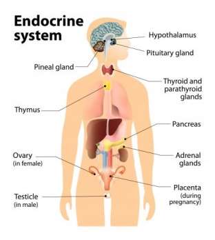 BPA may change the DNA structure in the body. It adds methyl groups into the DNA that stops the DNA expressions. However, it may have different responses varying on the dose of BPA and does not respond in a similar way to BPA as the classic receptors. It can also cause reproductive disorders such as effecting the egg maturation, cause small increases in levels of testosterone in the blood of sex hormones in men, breast cancer, and asthma occurring at the same time as BPA production that is common in the increase of asthma rates.
Fetal Development
Fetal development is the process in which a human embryo and later fetus develops during pregnancy, from fertilization to birth.
BPA in maternal urine may damage fetal growth [5]. Levels of BPA in urine between 125 pregnant women during the first three months were inversely associated with fetal head circumference in the third three months [14]. In a study population of 339 women and children, higher urinary BPA concentrations in the third trimester of pregnancy were related with somewhat higher birth weight in offspring, but were not associated with head circumference [14]. There is a positive correlation between the BPA concentration of maternal urinary and birth weight and head circumference in the study of 191 children and women [14]. Most urine studies are found higher BPA concentrations in children than adults [8].
A bottle-fed baby will have much higher levels of BPA than a breastfed baby [8]. Breast milk often holds BPA of lower levels due to the mother’s exposure from canned foods and other assets.
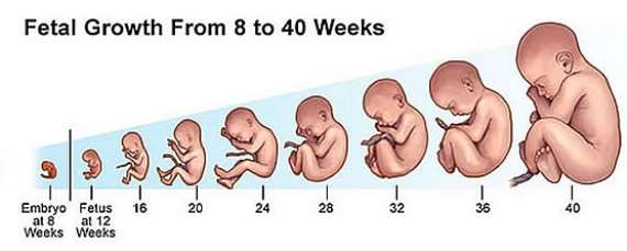
Changes that Occurred in Pregnancy When Exposed to BPA
In pregnant women, BPA exposure affects not only the mother, but also the fetus growing inside of the mother.
Maternal and fetal exposure to BPA was linked with disorder of thyroid function throughout pregnancy and the newborns are characterized by a decrease in circulating thyroxine levels [18]. “BPA concentrations in the mother blood in this experiment were fluctuating between injections from 15 to 1 time the highest blood levels reported in pregnant women in the literature,” notes Viguié [18].
Changes can also include brain growth. As neurons start growing, chloride levels in the cell are important for directing their growing nerves to their appropriate place in the brain. If chloride levels are high, the circuits do not form and connect correctly. Exposing human nerve cells to BPA, researchers found out that the chemical prevents KCC2 (Potassium chloride transporter member 5) from its functioning in lowering the levels of chloride [20]. Sex hormones are contributed in brain development. Hormones play a key role in the process of a determining phase in which the neurons are involuntary, brain regions develop, and synapses begin to control together the outcome society of cells. For example, signals provide the order of the period and sequence of neuronal development.
Researchers have found out that mothers during pregnancy with higher levels of BPA in their urine tend to have 3-year-old girls with more of an anxious and depressed behavior and have less emotional control [6]. Mothers with higher levels of BPA were not shown in boys and had no affect on their behavioral and emotional control [6]. There could be an increase in childhood disorders such as clinical anxiety and hyperactivity [11]. The development in the pituitary glands are also important because it is composed of hormone-producing cells which is significant for homeostasis and reproduction.
Human Health Effects Assessment [21/22]
An oral doing study in rats result in that the tissue concentrations of BPA-derived radioactivity were highest in the liver, kidney, carcass and lowers in brain and testes. There was no big of a difference between adult and neonatal animals.
Low levels of BPA were found to be spread in the foetus after a high oral dose of BPA in pregnant rats. The spread of BPA to the foetus (or placenta) did not modify the overall pharmacokinetic (movement of drug in the body) chance of BPA in pregnant rats, compared to non-pregnant rats.
Studies in rats revealed that in newborns the glucuronidation passageway is more open to capacity than in adults that implies a higher inner exposure to free BPA. Due to the high water solubility, BPA glucuronide produced during first-pass metabolism speedily clear from human blood by the kidneys and emitted in the urine with life-threatening half-lives of around 5 hours after oral supervision.
There is a main species difference between humans and rodents in the removal of BPA glucuronide from liver. In differentiation to humans, BPA undergoes enterohepatic circulation in rats, which effects in slow elimination and increased total availability of free BPA in rodents.
Current data described; however, informed the discovery of free BPA concentrations in human and rodent blood and urine samples and advise that there might be higher human exposure to free BPA.
Evidence on internal exposure to BPA is of meaning because toxic effects are related to blood concentrations relatively than to outer exposure and that the evidence would also allow the finding of species differences in metabolism. Free BPA was discovered in human plasma and urine in several studies.
BPA is measured to be quickly conjugated into BPA glucoronide and BPA sulphate and thus eliminated from the human body because the water solubility of the metabolites. Internal exposure to free BPA existing for biological activity within the body is then expected to be very low. Data from measurements of un-conjugated (free) BPA in human blood and urine; nonetheless, indicate higher internal exposure of humans to free BPA. Newborns are likely to be exposed to higher internal BPA standards due towards immature glucuronidation activity.
Reproductive Toxicity of BPA
Reproductive toxicity of BPA is the key importance for risk evaluation. A significant number of studies on the reproductive toxicity of BPA in experimental animals are covering effects at very low doses, such as prostate growth and development, early puberty, body weight, etc.
Some research studies have designated a non-monotonic dose response curve with effects on reproduction and development at very low dose levels (µg/kg bw-range) declining at intermediate levels, with different effects appearing at higher mg/kg-bw doses. NOAELs (no-observed-adverse-effect-level) since such studies would be at several orders of magnitude lower than the TDI (tolerable daily intake) and lower than measured exposure levels in humans. However, these studies have organizational limitations and were often not using the (relevant) oral way of direction. Recommendation studies in acquiescence with GLP (Good Laboratory Practice), using the oral way of exposure, did not approve the above stated low dose results of Bisphenol A on reproduction and maturity.
BPA Exposure Can Lead to Miscarriages [18]
Phthalates are a group of chemicals used to make plastics more durable and flexible, such as beauty products, soaps, and children’s toys. According to Centers for Disease and Control and Prevention, people can be exposed to phthalates by eating and drinking from containers containing the chemical. Phthalates have been connected to health issues such as the sexual development of males and attention-deficit disorder in children.
One study observed 501 couples that were trying to become pregnant. The couples in the study were cross-examined and surveyed and provided urine samples to measure their BPA and phthalate levels. Researchers discovered that men, not women, with high phthalate concentrations experienced a 20% failure in fertility and took longer to get their partners pregnant than men with lower phthalate concentrations.
American Society of Reproductive Medicine President Linda Giudice said that phthalates interfere with the hormone systems, known as endocrine disruptors.
” “Phthalates are anti-androgens, and other studies have shown that environmental levels of phthalates in infertile men correlate with increased rates of sperm DNA damage, low sperm counts and abnormal sperm,” she said.”
114 women were requested to give blood samples four or five weeks into their pregnancies in a smaller study. The blood samples were not examined until after the 114 women gave birth to live babies or after they had a miscarriage if it happened in the first semester. 68% of the pregnancies ended in miscarriages.
Researchers established that women of high levels of BPA in their blood were in the highly risk of miscarriage compared to women of low levels of BPA. They believed that there should be more studies on how the chemical affects pregnancy.
Case Study #1 [13]
In this case, the single and unemployed mother was a 26-year-old African-American woman with three reported past pregnancies and only one living child. She had no important past medical history and negative prenatal lab testing. She had a 27-week urinary BPA concentration of 583µg/g creatinine (1,250 µg/L), which was the highest BPA value recorded within the entire group that had n=389 participants and 1,100 urine samples. Her prenatal urinary BPA concentrations were respectively at 16 weeks and a birth were at the 84th (4.1 µg/g) and 49th (1.9 µg/g) percentiles. The case had a follow up with the CDC National Environmental Health Laboratory because of the concerns of the 583µg/g urinary BPA concentration could be a false outcome. The CDC established this result by extracting the sample twice to conduct repeat testing. The test values were precisely close to the original value and higher than the highest standard on the calibration curve. They had also acknowledged that most BPA in the urine was conjugated; therefore, it didn’t reflect external contamination. The case mother had concentrations of serum cotinine, blood lead, and urinary phthalates varying from the 25th to 84th percentiles in the group.
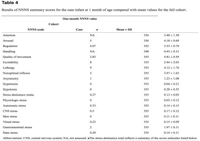
The male infant was born in 2004 (39 weeks of development) by vaginal delivery without having complications. The infant had a normal course in the newborn nursery and a normal NNNS (Neonatal Network Neurobehavioral Scale) at 14 hours and was discharged from the hospital after about 38 hours of birth.
At the 1-month neurobehavioral assessment, the infant showed the following clinical signs or indications: hyper tonicity in the trunk and upper and lower extremities, tremors, high-pitch cry, extreme irritability, etc. The infant had high NNNS scores for excitability, laziness, and stress self-denial and lower scores for instruction and quality of movement.
In order to determine the potential sources of the higher urinary BPA concentration and to keep up on the development of the child, the case used the prenatal survey and conducted a focused continuation telephone interview with the mother in fall of 2009. She reported drinking five canned soda beverages per week in the month before completing her prenatal survey at 22 weeks of development. The mother’s diet consisted of canned or frozen vegetables one to three times per week and fresh vegetables four to six times per week. The mother informed eating canned ravioli every day during the period that the 26-week urine sample was taken. She had daily exposure to plastics that included heating and using plastic food storage containers in the microwave and eating and drinking from plastic plates and cups. She was jobless throughout the pregnancy and living with her 4-year-old daughter during the time she was pregnant with the case infant. The mother’s home was within 2 miles of a whiskey factory, but there were no other industrial buildings in close proximity. Concerning her child’s medical condition, she stated that her primary care physician monitored her child with normal checkups and did not note any abnormal behavioral or developmental findings or other health problems. The mother reported the child was in school and performing well.
This case study emphasizes a high prenatal urinary BPA concentration at 26-week development and a peculiar neurobehavioral examination at one month of age. It is likely that many causes of BPA exposure had subsidized to the mother’s high urinary concentration during pregnancy. This case increases the possibility that gestational BPA exposure may be correlated with strange infant behavior, since BPA is a supposed neurotoxicant that has caused temporary bizarre results in toxicological studies.
Case Study #2 [16]
A case study called “The Generation R” wants to confirm the relation of prenatal BPA exposure with fetal growth by measuring at birth and urinary samples.
“The Generation R” study is a population-based potential group study of growth, development, and health from early fetal life until young adulthood in the Netherlands. Pregnant women with a likely delivery date between April 2002 and January 2006 were invited to participate in the study. 9,778 pregnant women participated in the study with 8,880 pregnant women and 898 women at birth of their child. General evaluation was carried out during early pregnancy (gestational age < 18 weeks), mid-pregnancy (gestational age 18-25 weeks), and late pregnancy (gestational age > 25 weeks), with biological samples.
219 women with 419 urine samples were available, with 26% of samples from first trimester, 28% from second trimester, and 46% from third trimester of pregnancy. All urine samples (65 mL) were collected between February 2004 and November 2005. All samples were taken between 0800 and 2000 hours in 100-mL polypropylene urine collection containers that were kept in a 20 hour in a cold room (4°C) before being frozen in 20-mL portions in 25-mL polypropylene vials at -20°C. The urine samplings were analyzed for BPA using cycle mass spectrometry.
Researchers used second- and third-trimester fetal ultrasound measurements with measurements of fetal size at birth to estimate growth rates during pregnancy. They measured growth characteristics to the nearest millimeter using consistent ultrasound procedures in the second (median, 20.5; range, 18.2-25.0 weeks) and third (median, 30.2; range, 27.4-33.8 weeks) trimesters. First-trimester measurements were used to start gestational age because the use of the last menstrual period for pregnancy dating has several limitations, and most women (76%) in our study population did not know the exact date of their last menstrual period. They used important-rump length for pregnancy dating until gestational age of 12 weeks, and bi-parietal diameter for pregnancy dating after in all women. Estimated fetal weight (EFW) was calculated using the formula. The intraclass correlation coefficient of fetal growth measurements was 0.95, based on 21 subjects, representing a strong relation for different fetal biometry measurements between and among observers. Internal indication curves based on the total Generation R group were made for fetal weight and fetal head circumference during pregnancy, showing typical parabolic patterns.
The discoveries from this group study supports the researcher’s hypothesis that higher concentrations of creatinine-based BPA in prenatal urine may lead to lower fetal weight and head circumference. Estimated relations between BPA and fetal growth were stronger and statistically substantial when three BPA measurements were measured instead of a one-time BPA measurement.
The strength of this study is the population-based approach with enrollment during the prenatal period and analysis of several urine samples. Another strength of this study is the numerous observations on fetal growth as well as repetitive measurements of BPA per subject. A restriction is the selective participation at standard, with mothers of lower socioeconomic status less represented in the study population.
The results imply that increased concentrations of BPA in urine during pregnancy are correlated with a decreased fetal growth for both fetal weight and head circumference. Moreover, this study supports the use of multiple measurements per subject to measure exposure in studies on exposure-response relations. The need for further proof before concluding that in the general population, BPA during pregnancy negatively affects fetal growth because earlier studies show contradictory results and the characteristic boundaries of the study.
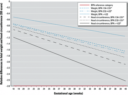
* Relative differences in standard scores for fetal weight and head circumference in various BPA exposure groups.
Case Study #3 [17]
In this case study, scientists measure the contamination of BPA in numerous kinds of human biological fluids by a unique enzyme: linked immunosorbent assay (ELISA).
Instead of taking urine samples from pregnant women, the researchers used blood samples from 30 healthy premenopausal women, 37 women with early and full-term pregnancy, and 32 umbilical cords at full-term delivery. Thirty-six ovarian follicular fluids were aspirated during IVF procedures. 32 and 38 amniotic fluids were taken by amniocentesis at 15-18 weeks development (early pregnancy) and by amniotomy (artificial rupture of membranes) at full-term C-section (late pregnancy) correspondingly. No chromosomal defects or major differences were found in the fetuses. The Kruskal-Wallis test, completed statistical studies between the groups. P < 0.05 was selected to signify a statistically meaningful difference.
According to the study, BPA was existent in serum and follicular fluid at 2.0 ± 0.8 (mean ± Standard Deviation; non-pregnant), 1.5 ± 1.2 (early pregnancy), 1.4 ± 0.9 (late pregnancy) and 2.4 ± 0.8 ng/ml (follicular fluid), as well as in fetal serum (2.2 ± 1.8 ng/ml) and amniotic fluid, verifying way through the placenta. There was no significant correlation between the maternal and fetal serum concentrations, signifying that BPA might be somewhat metabolized in the fetus. There was approximately a 5-fold difference in BPA concentrations between amniotic fluid found at 15-18 weeks development (8.3 ± 8.9 ng/ml) and other fluids (P < 0.0001). Yet, amniotic fluid levels declined drastically at phase (1.1 ± 1.0 ng/ml) and no longer changed from the levels of other fluids.
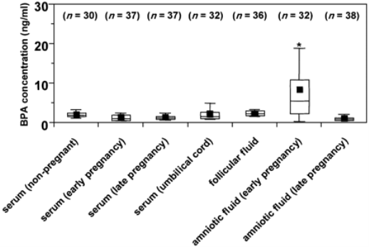
* Bisphenol A (BPA) concentrations in human biological fluids. Data is presented as box and whisker plots, where boxes hold values between 25th and 75th percentiles, horizontal lines symbolize median values, and ‘whiskers’ give the 95% range of the values. Plots (â-ª) represent mean values. Number of samples shows in parentheses. *P < 0.0001 compared with other biological fluids.
Scientists compared BPA values found by their ELISA to the established reverse-phase high-performance liquid chromatography (HPLC) analysis in order to verify the results of their ELISA for BPA in the amniotic (inner most membrane that encloses the embryo) fluid samples.
The results showed a positive linear correlation between the two procedures. The source of amniotic fluid in the first half of pregnancy likely is the passage of serum or other body fluid through the membranes or tissue surfaces of fluid from maternal plasma across the membranes that protect the placenta and the cord because in structure, the fluid is almost identical to a transudate of plasma. In the second half of pregnancy, there is a gradual mixture of fetal urine. From this idea of assessment, in the first half of pregnancy BPA is moved from maternal plasma to amniotic fluid and stored in the uterine cavity because of the low metabolic allowance of BPA. Therefore, a fetus is exposed to a large amount of BPA. Higher levels of BPA in the mid-term amniotic fluid show accumulation and the following significant exposure that may be clarified by immature liver function including UGT (uridine diphosphonate glucuronosyl transferase) activity. In the second half of pregnancy, a fetus can gulp a large amount of amniotic fluid and conjugate BPA alongside with the maturation of liver function. Because BPA presumably returns through placenta from fetus to mother or is weakened by fetal urine, BPA levels in amniotic fluid may decrease at time.
There is much to be explained about the connection of early BPA exposure in the recent occurrences in humans, for instance, increased genital abnormalities in boys, earlier sexual maturation in girls, decreased sperm count in men, and increased breast cancer in women.
Reversing/Reducing BPA Toxicity [1]
There are many ways of reversing and reducing BPA toxicity. Fermented foods such as kimchi, natural sauerkraut and kefir contain probiotics. Black tea is found to reduce BPA toxicity. It contains quercetin which is a flavonoid found in fruits, veggies, leaves, tea, and whole grains. Sweating can also reduce the toxicity. Researchers discovered that BPA was found in sweat so induced sweating, is a possible method for BPA elimination. Besides the methods above for possible elimination of BPA, the lower use of canned foods and plastic containers can reduce BPA toxicity.
FDA’s Point of View/Research [9/7]
The FDA (Food and Drug Administration) continues to approve BPA’s safe use as a food-packaging compound base on the ongoing safety review. According to the FDA’s research on BPA’s safety, it is confirmed to be harmless. The FDA confirmed that BPA is below safe limits set by the government agencies and that BPA doesn’t stay in the body forever, we process it and eliminate it very quickly. The abandonment of the amendment of the food additive regulations was not based on safety, but based on the no longer necessary for the use of food additives. The safety of food additive is not important to the FDA’s purpose concerning whether a specific use of the food additive has been discarded. It is said that the FDA will continue to support the safety of BPA and will continue to update the public on new important information when available.
The FDA advises the public against putting boiling, hot liquid in plastic containers made with BPA if planned to drink the water [12]. BPA levels are high in food when containers or items are made with the chemical are heated and come in interaction with the food [12]. Therefore, the FDA does not allow BPA in plastic that is used to make baby bottles or toddler cups [12].
Conclusion
According to the research, there is a correlation between BPA and fetal development. BPA can affect the internal developments and lifestyle, particularly in pregnant women. The FDA no longer provides the use of BPA-based epoxy resins and polycarbonate resins as coatings in packaging for infant formula and in baby bottles/sippy cups because there is no longer use of food additives. The result of case study #1 and #2 verifies studies authenticating numerous sources of BPA exposure in humans. In case study #1, those discoveries can inform health car practitioners that the general population may be exposed to high concentrations of BPA during maturity that is a serious time for organ system and brain development. In case study #2, the results proposes that higher concentrations of BPA in urine during pregnancy are associated with a diminished fetal growth for both fetal weight and head circumference. The outcomes of these risk assessment depend on how an amount of crucial causes are considered, for example the approximations of exposure levels. In case study #3, it concludes that the fetus does intake a great amount of BPA through the mother. However, there is much to be clarified about the connection of initial BPA exposure in the recent events in humans.
